So today we're gonna focus on the lungs. I think we are all familiar with how to scan for pneumothorax and for pleural effusions/hemothorax as part of the Extended FAST exam, but what about pathology within the lungs themselves? I know what you are thinking, "Duran, I thought you said that we can't see the lungs on ultrasound. They're full of air." You are correct, however, if the lungs are either surrounded by fluid or if they are fluid filled then you can see them on US. So that brings us to advanced lung ultrasound or the A's and B's of Lung US.
Let's start with A or A-lines. A lines are a normal finding on lung US and you have probably seen them before and not really paid much attention to them. They are a reverberation artifact.
Notice that each line is equally spaced? Notice also that they do not continue all the way down the screen. This occurs because the sound wave is bounced between two reflectors. Each time it hits the reflector it bounces a portion of the sound beam back to the machine. Eventually the entire sound beam is consumed.
So the appearance of A-lines on lung US is completely normal and should be expected.
On the other hand, B-lines are not normal and indicate "intraparenchymal lung fluid." Huh? The lung is normally full of air, but in pathologic states, it become like a sponge and the alveoli become full of fluid. US can't tell if the fluid is pus as in a pneumonia, blood as in a pulmonary contusion or extracellular fluid as in pulmonary edema. It can just tell you that the lung parynchema is full of fluid. It is up to you as the clinician to determine the cause. So what does intraparenchymal fluid look like?
At it's simplest it appears as B lines. B lines are comet tail artifacts that begin at the pleura and continue to the end of the screen.
Seeing 3 or more of these B lines in a single rib interspace is pathalogic, but they often present as a curtain or spotlight moving accross the screen as the patient breaths.
video
When the lung becomes completely full of fluid, such as in severe pulmonary edema or severe pneumonia, the lug will start to resemble liver tissue. Notice in the picture below how much the lung and the liver look alike. The presence of a pleural effusion make it easier to see the lung as it surrounds the lung, increasing visualization.
So how do we scan the lungs efficiently to look for fluid within the lungs themselves?
The Lung Scan Protocol
The protocol for scanning the chest for intraparenchymal fluid is a
little more extensive and time consuming than the E-FAST scan we do for
pneumonthorax. This protocol requires that we divide each side of the
chest into 5 segments: 2 anterior, 2 lateral and 1 posterior. The anterior and lateral chest are each divided into two segments: upper and lower.
Each individual segment must be scanned in order to accurately rule out fluid. Therefore, there should be a picture from each segment submitted for this protocol- meaning 10 pics total as both sides need to be scanned.
There have been several articles in the literature recently dicussing the use of ultrasound prior to even plain CXR for the diagnosis of pneumonia. Although these studies show good results in that US was able to accurately determine pna, I am not sure that it will replace the good old CXR. Interestingly enough pneumonia can appear as B lines or if it is large enough, air bronchograms may be visualized on US! Amazing, right? Here are a couple of pics taken from these articles illustrating air bronchograms on US. Check out these articles if you get a chance or scan your next patient with pneumonia and see if you can see this.
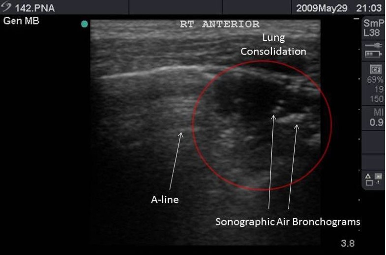
Prospective application of clinician-performed lung ultrasonography
during the 2009 H1N1 influenza A pandemic: distinguishing viral from
bacterial pneumonia.Tsung JW1, Kessler DO, Shah VP.Crit Ultrasound J. 2012 Jul 10;4(1):16. doi: 10.1186/2036-7902-4-16.
Is lung ultrasound superior to CT? The example of a CT occult necrotizing pneumonia. Lichtenstein D1, Peyrouset O2. Intensive Care Med. 2006 Feb;32(2):334-335. doi: 10.1007/s00134-005-0004-6. Epub 2006 Jan 27.16468074 doi:10.1007/s00134-005-0004-6
The dynamic air bronchogram. A lung ultrasound sign of alveolar consolidation ruling out atelectasis. Lichtenstein D1, Mezière G2, Seitz J3.Chest. 2009 Jun;135(6):1421-1425. doi: 10.1378/chest.08-2281. Epub 2009 Feb 18.


